Next-Generation Material Discrimination for Food Inspection
Sens-Tech’s latest Dual-Energy TDI innovations are setting a new benchmark in food inspection, enabling reliable material discrimination at industrial processing speeds.
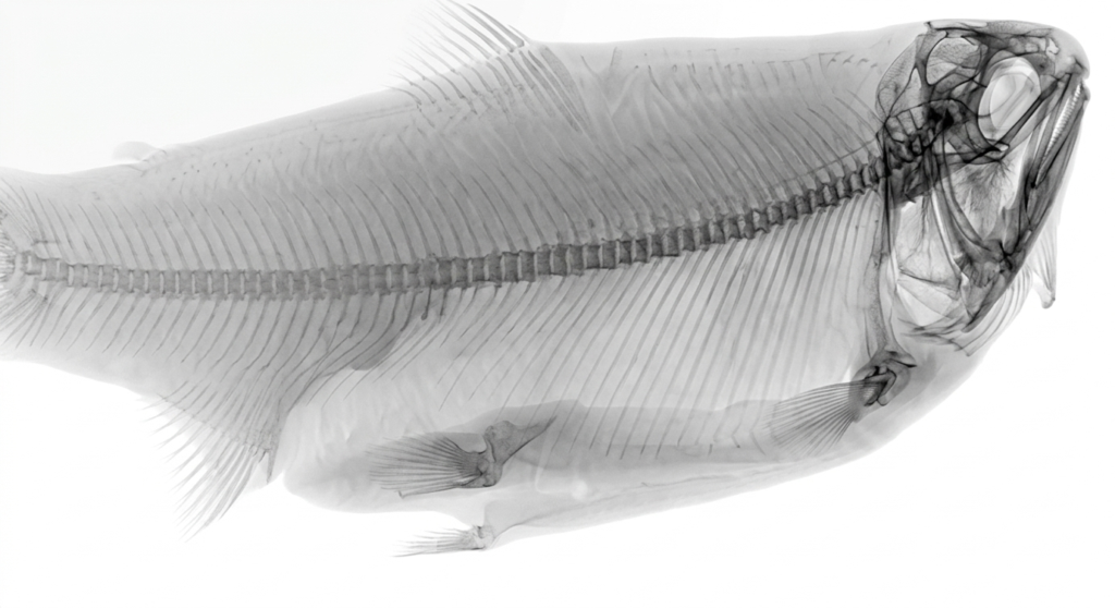
The Challenge
Tiny fish bones with varying densities are notoriously difficult to detect and remove. As a result, seafood processors often spend significant time and resources identifying them manually. Conventional detection technologies can help, but they frequently lack the accuracy needed for high-speed production environments.
Limitations of Existing Technologies
Small-pixel single-energy TDI cameras may highlight bone-like structures, but without dual-energy imaging they cannot reliably confirm whether the detected material is actually bone.
More advanced photon-counting arrays can offer improved image quality, but they are often costly, less durable, and slow to integrate—making them impractical for high-throughput industrial applications.
The Sens-Tech Solution
Sens-Tech’s Dual-Energy TDI technology overcomes these limitations. Our unique innovations enable seafood processors to:
- Detect more contaminants with greater confidence
- Reduce product waste
- Maintain high-speed processing efficiency
Download our white paper below to find out how we do this!