
Inline Detection of 0.3 mm Fish Bones Using Low-Energy X-ray TDI Imaging – Why Sub-Millimeter inspection Is a Platform Stability Challenge — Not a Resolution Specification
Detecting 0.3 mm fish bones inline at industrial conveyor speeds represents a boundary condition problem in food inspection X-ray. At this scale, contrast is limited, photon statistics become critical and mechanical stability directly impacts detectability.
Below we demonstrate detection feasibility using:
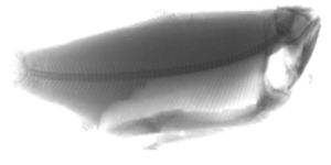
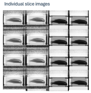
Individual slice images and low/high-energy TDI reconstructions. These were evaluated to determine signal formation, SNR limits and achievable detection performance.
Detecting sub mm fish bones inline is often framed as a detector resolution problem.
It is not.
Inspection set up
Source Parameters
- Tube Voltage: 50 keV
- Tube Current: 4.5 mA
- Source Distance: ~800 mm
Acquisition Parameters
- Conveyor Speed: 0.5 m/s
- Detector Pitch: 1 mm × 1 mm
- Integration Time: 2000 µs
- 8 TDI rows
- Gain: 9.375 pC
At this scale, detection becomes a system-level signal integrity challenge governed by:
- Photon statistics
- Signal chain stability
- Mechanical precision
- Calibration repeatability
- Scatter control
OEMs who approach this as a pixel-size specification exercise typically encounter unstable performance in production.
The Physics Reality
Fish bone is not metal.
It is a low-Z, calcium-based structure embedded in soft tissue with only marginal attenuation contrast at 50 keV.
At 0.3 mm thickness, the expected differential attenuation between bone and surrounding muscle produces only a ~1–2% signal variation per pixel.
That means:
The system must operate with effective SNR levels above ~50–80 to reliably separate bone from noise in real production conditions.
This is not a nominal imaging task but is a precision signal detection task.
At line speeds of 0.5 m/s, motion during one integration equals exactly one pixel (1 mm). TDI accumulation across 8 rows improves SNR by ~√8 ≈ 2.8×.
At 0.3 mm thickness, attenuation contrast in soft tissue is small. Detection is therefore limited by:
- Quantum noise
- Scatter contamination
- Detector stability
- Motion blur
- Electronic noise floor
The task becomes a signal-to-noise engineering problem, not a nominal resolution problem.
Strategic Takeaway
At sub-millimeter detection limits:
Photon counting is not a silver bullet.
Detection margin is primarily governed by:
- Photon statistics
- Stability
- Scatter management
- Calibration precision
Photon counting shifts noise composition — it does not change the fundamental √N limit.
If the platform is already quantum limited, architecture choice becomes a trade-off between:
- Flux handling
- Stability
- Cost
- Complexity
- Scalability
A single slice is insufficient for stable 0.3 mm detection.
TDI Reconstruction
After 8-row accumulation:
- Noise is reduced.
- Bone contrast becomes perceptible.
- Stability improves significantly.
However:
The detection margin remains narrow.
Any instability directly impacts detectability.
Detector Gain Setting (9.375 pC)
High gain improves sensitivity but risks:
- Reduced dynamic range
- Saturation in thick regions
- Amplified electronic noise if frontend not optimized
Detection of 0.3 mm bones requires:
- Low electronic noise floor
- Stable charge integration
- Precise gain calibration across rows
Individual Slices
- Slice-level images operate near the quantum noise floor.
- Row-dependent gain variations are visible.
Structured noise and scatter are present.
Detection is quantum-noise dominated.
Photon counting would:
- Remove electronic noise floor
- Offer modest scatter suppression
- Introduce count-rate management complexity
Expected improvement in 0.3 mm detectability:
Likely 10–25% margin increase — not 2×.
Increasing TDI rows from 8 to 16 would produce similar or greater improvement with lower system risk.
Conclusion:
- Low energy acquisition is required for sub-millimeter bone detection.
Inline detection of 0.3 mm fish bones at:
- 50 keV
- 0.5 m/s
- 1 mm pitch
- 8-row TDI
is technically feasible but operates at the edge of SNR limits.
Sub-millimeter bone detection is not a component problem.
It is a platform integrity problem.
At 50 keV with 8-row TDI:
- The physics allows detection.
- The margin is narrow.
- Stability determines commercial success.
The real differentiator is not resolution.
It is whether the system can deliver the same SNR — every shift, every day, every installation.
For more information, contact
Paul Hurtado, Head of Sales and Marketing at Sens-Tech.